Here are some brief updates, which won’t impact any of you on the list serve (please note a virtual-only talk on 4/1 Wed).
Author: Elisa Medeiros
BIRC’s response to the Coronavirus Outbreak Update #6 3-16-20
With many states and counties advancing into lockdown (SF is stating lockdown of the whole city together with neighboring counties effective tonight – just announced! I believe it will come our way to CT sooner than we think/hope. I hope I am wrong), we have made some adjustments.
We technically remain open until the university announcement, but …
RESEARCH SCANS
We have cancelled all research scans until 4/6 and will continue to discourage scanning unless you can convince us 🙂
CLINICAL SCANS
While UCHC has decided to only cancel elective surgery at this point, and no outpatient services are cancelled, we have agreed to cancel/postpone all clinical scans as well. We will scan the remaining patients if there are critical needs but have suspended all clinical scans at least until 4/6 as well. We are currently not booking any new patients. Thanks for Leo Wolansky (UCHC Radiology Department Chair)’s understanding in the service of protecting our staff.
Hence no disinfection will be done as complete telecommuting has started.
If you have critical needs please contact me.
Stay safe and happy…
Fumiko
BIRC’s response to the Coronavirus Outbreak Update #5 3-16-20
A quick update to you all.
RESEARCH SCANS
We are currently technically open. But we do not have any research scans until the weekend (and they are non-EEG) and will likely close as soon as we get guidance from VPR. They are in discussions and will propose recommendations to the President this afternoon. We are urging the university to stop all non-critical/essential research that does not impact human health ASAP. We should have updates before any further research scans are done.
CLINICAL SCANS
We are in discussions with UCHC Radiology to stop clinical MRI as well as they are non-priority scans.
OTHER ACTIVITIES
We remain open virtually for any consultations or training that can be done remotely. We will suspend in person MRI safety training until further notice. LMK if this impacts your research plans.
Please do not come in to use the conference rooms or data room unless necessary. While we have staff telecommute, we will not be disinfecting the areas as we had originally planned and was doing last week. If you do need to come in to BIRC, please contact me.
Thank you,
Fumiko
BIRC’s response to the Coronavirus Outbreak Update #4 3-13-20
At this time BIRC will continue to be fully operational. Any scheduled research will not be impacted, and investigators can schedule additional research times as needed.
Please note that we will be complying with the University’s request to have staff and faculty telecommute when possible and may not be in the facility during normal business hours; however, please feel free to email any questions to Fumiko Hoeft, Roeland Hancock, or Elisa Medeiros.
Scheduling Seed Grant Studies During Spring 2020 Semester
MRI Scanner Operation Training for Qualified Candidates
The Brain Imaging Research Center now offers qualified candidates the opportunity to learn how to operate the Siemens Prisma 3T MRI Scanner to perform brain research studies. This training will consist of three components:
-
-
- Post doc with a commitment to remain for a minimum of one year (must be endorsed by PI)
- Graduate student who has completed their Masters degree and must be endorsed by PI
- Formal knowledge of basic MRI physics
- Completion of Level 1 and Level 2 Safety Training
- CPR certified (must provide documentation prior to scanning humans)
-
Online classes available at redcross.org/take-a-class/online-safety-classes
-
-
- PI name, duration of contract, and written endorsement
- Proof of formal basic MRI physics education
- Any previous MRI experience
- Study name, projected start date, and expected number of participants
-
-
-
- Application submission: October 7-October 18 2019
- Candidate acceptance notification: October 25 2019
- Didactic and Instrumentation training: November 2019 (dates TBD)
- Scanner Operation: November 2019 until completed
-
Professors Myers and Eigsti Receive Five Year Training Grant
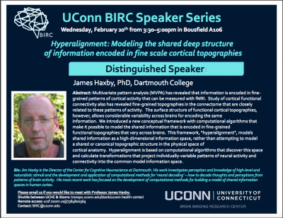
Talk: Dr. James V. Haxby, Dartmouth College
Dartmouth College
Distinguished Speaker
Wednesday, February 20 2019 3:30-5:00PM Bousfield A106
Abstract: Multivariate pattern analysis (MVPA) has revealed that information is encoded in finegrained patterns of cortical activity that can be measured with fMRI. Study of cortical functional connectivity also has revealed fine-grained topographies in the connectome that are closely related to these patterns of activity. The surface structure of functional cortical topographies, however, allows considerable variability across brains for encoding the same information. We introduced a new conceptual framework with computational algorithms that make it possible to model the shared information that is encoded in fine-grained functional topographies that vary across brains. This framework, “hyperalignment”, models shared information as a high-dimensional information space, rather than attempting to model a shared or canonical topographic structure in the physical space of cortical anatomy. Hyperalignment is based on computational algorithms that discover this space and calculate transformations that project individually-variable patterns of neural activity and connectivity into the common model information space.
Research Focus: My current research focuses on the development of computational methods for building models of representational spaces. We assume that distributed population responses encode information. Within a cortical field, a broad range of stimuli or cognitive states can be represented as different patterns of response. We use fMRI to measure these patterns of response and multivariate pattern (MVP) analysis to decode their meaning. We are currently developing methods that make it possible to decode an individual’s brain data using MVP classifiers that are based on other subjects’ data. We use a complex, natural stimulus to sample a broad range of brain representational states as a basis for building high-dimensional models of representational spaces within cortical fields. These models are based on response tuning functions that are common across subjects. Initially, we demonstrated the validity of such a model in ventral temporal cortex. We are working on building similar models in other visual areas and in auditory areas. We also plan to investigate representation of social cognition using this same conceptual framework.
Visitors from UCHC are encouraged to use the UCHC-Storrs shuttle service. Talks can also be joined remotely. Please contact us if you are interested in meeting with the speaker.

UConn BIRC Trailblazer Award Announced
Issue Date
December 19, 2018
Background
Since the opening of the University of Connecticut (UConn) Brain Imaging Research Center (BIRC) in June 2015, there has been an increase and diversification of user-base, neuroimaging-related extramural grants, and neuroimaging expertise of students and faculty. However, there is still room for greater utilization of BIRC, which presents opportunities for BIRC to offer the resources to perform high-profile and neuroimaging-intensive research that other fully occupied imaging centers cannot offer.
Objective
The BIRC Trailblazer Award was created to allow research teams to perform cutting-edge research and/or perform research that will benefit the BIRC community at-large. The objective of the 2019 BIRC Trailblazer Award is to fund: (1) high-risk high-reward projects with exceptional innovation that lead to raising the visibility of UConn, College of Liberal Arts and Sciences (CLAS) and BIRC; and/or (2) projects that will benefit the BIRC community at-large (e.g. methods development). The project is intended to lead to high-profile peer-review publications, release of a public database, and/or work that is cited and utilized by large-number of UConn researchers in their grants and manuscripts. The project should also lead to large-scale and high-profile extramural grant applications shortly after the end of the funding period.
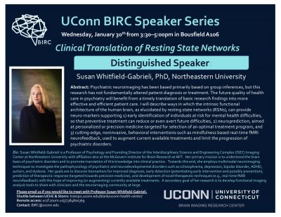
Talk: Clinical Translation of Resting State Networks
Northeastern University and MIT
Distinguished Speaker
Wednesday, January 30 2019 3:30-5:00PM Bousfield A106
Abstract: Psychiatric neuroimaging has been based primarily on group inferences, but this research has not fundamentally altered patient diagnosis or treatment. The future quality of healthcare in psychiatry will benefit from a timely translation of basic research findings into more effective and efficient patient care. I will describe ways in which the intrinsic functional architecture of the human brain, as elucidated by resting state networks (RSNs), can provide neuro-markers supporting 1) early identification of individuals at risk for mental health difficulties, so that perventive treatment can reduce or even avert future difficulties, 2) neuroprediction, aimed at personalized or precision medicine targeted for selection of an optimal treatment program, and 3) cutting-edge, noninvasive, behavioral interventions such as mindfulness based real-time fMRI neurofeedback, used to augment current available treatments and limit the progression of psychiatric disorders.
Bio: Susan Whitfield-Gabrieli is a Professor of Psychology and Founding Director of the Interdisciplinary Science and Engineering Complex (ISEC) Imaging Center at Northeastern University with affiliation also at the McGovern Institute for Brain Research at MIT. Her primary mission is to understand the brain basis of psychiatric disorders and to promote the translation of this knowledge into clinical practice. Towards this end, she employs multimodal neuroimaging techniques to investigate the pathophysiology of psychiatric and neurodevelopmental disorders such as schizophrenia, depression, bipolar disorder, ADHD, autism, and dyslexia. Her goals are to discover biomarkers for improved diagnosis, early detection (potentiating early intervention and possibly prevention), prediction of the therapeutic response (targeted towards precision medicine), and development of novel therapeutic techniques (e.g., real-time fMRI neurofeedback) with the hope of improving (or augmenting) currently available treatments. A secondary goal of her research is to develop functional imaging analysis tools to share with clinicians and the neuroimaging community at large.
Visitors from UCHC are encouraged to use the UCHC-Storrs shuttle service. Talks can also be joined remotely. Please contact us if you are interested in meeting with the speaker.
