The UConn Brain Imaging Research Center is located in the David C. Phillips Communication Sciences Building (PCSB; directions) on the Storrs campus. BIRC offers the resources for nearly any MRI, EEG or neuromodulation study. Please submit the study request form prior to beginning any project and complete any required training before using the equipment.
A template Facilities and Resources page is available. If you will be submitting a grant that uses BIRC resources, please contact us.
Magnetic Resonance Imaging
Siemens Prisma 3 Tesla scanner equipped with a range of head and body coils:
- 20-channel head/neck coil
- 32-channel head coil
- 64-channel head/neck coil
- 32-channel spine coil
- Large and Small Shoulder coils
- Large and Small Flex coils
- Hand/Wrist coil
- Foot/Ankle coil
- Knee coil
- 18-channel body array coil
Product and research pulse sequences for:
- Structural imaging (Siemens or MGH sequences)
- Diffusion imaging (Siemens or CMRR sequences)
- BOLD
- Susceptibility Weighted Imaging (SWI)
- Spectroscopy and chemical shift imaging (Siemens or CMRR sequences)
- Simultaneous Multislice (SMS) EPI (Siemens or CMRR sequences)
- Arterial Spin Labeling
Stimulus presentation and response hardware for high fidelity auditory and visual presentation:
- Avotec Silent Scan audio system
- Optoacoustics OptoACTIVE II audio system
- Hyperion MRI Digital Projection System
- Current Designs MR-compatible response pads
- In-Scanner Eye Tracking (SR Research Eye Link 1000 Plus)
- Presentation using a PC, Mac, or your own laptop
Scanner QA
The scanner has been extensively validated in internal tests. We have replicated multiple visual field studies as well as functional localizers across scanners from multiple other sites. We perform QA tests every week using both ACR (American College of Radiology) and Siemens phantoms. In addition, we perform a scanner stability measurement from MGH. Results from these three weekly tests are automatically processed by routines at the BIRC and are monitored carefully for any sudden changes in the output. The scanner is remarkably stable, which we have attributed to both our diligent efforts at managing the magnetic field as well as regular service from Siemens.
EEG
BIRC houses two high-density (256-channel) Philips/EGI gel-free EEG systems. A 64-channel Brain Products actiCap system is available at CSSERL.
- 256-channel EGI NetAmps 400 EEG system for out-of-scanner use
- MR-compatible 256-channel EGI NetAmps 410 for simultaneous MR-EEG recording
- Sound-attenuated booth for EEG and behavioral experiments with visual and auditory presentation
- EGI Geodesic Photogrammetry System for electrode localization
- Complete range of electrode net sizes for child and adult friendly electrode application
- Physio16 box (MR compatible) for recording ECG or other physiological signals
- Cedrus StimTracker for precise event marker timing
Neuromodulation
BIRC houses a MagVenture MagPro X100 transcranial magnetic stimulation (TMS) stimulator with:
- Localite TMS Navigator system
- Biphasic and monophasic waveforms
- Flat and 120º butterfly coils
- Liquid-cooled active/sham butterfly coil
- Theta burst capability up to 100Hz
- 2-channel Biopac system for EMG recording
The EGI NetAmps 400 is also equipped with the GTEN neuromodulation package for high-definition transcranial electrical current stimulation (tDCS/tACS/tPCS). Please contact us if you are interested in using this system.
Experimental Control
Licenses for major experimental control softwares are available for use with MRI, EEG or behavioral protocols. These licenses may not be used outside of BIRC. Supported software includes:
- E-Prime Pro 2.0.10.356 (with legacy support for 2.0.10.353, 2.0.10.242)
- MATLAB/Psychtoolbox
- Presentation
- Experiment Builder
- PsychoPy 2 and 3
- OpenSesame
Interfacing with MRI
For the most part, all of these software products work the same inside of the scanner as they do outside of the scanner. However, there are some nuances that you will want to take into account. First and foremost, the scanner sends either a 5 or a t whenever the TR begins. This is relevant because you likely want to have your program begin after it receives the first TR marker. Your programs may use subsequent TR markers to synchronize with the scanner.
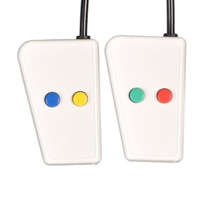
We have two response boxes and a joystick available to users (made by Current Designs). Only one of these devices can be used at a time. Button boxes are available in the 2x2 button layout
or straight 1x4 layout.
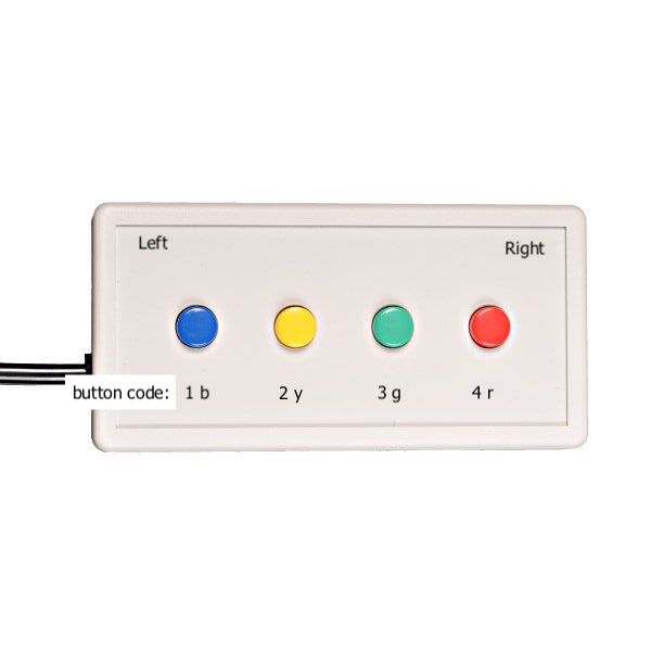
The readout of the buttons corresponds to either of the following sequences:
| Readout | Trigger Pulse |
| Blue = 1, Yellow = 2, Green = 3, Red = 4 | 5 |
| Blue = b, Yellow = y, Green = g, Red =r | t |
You will want to ensure that experimental control software accepts those as key presses. We recommend that you test these as much as possible before showing up for your development session. For Psychtoolbox users, the KbName of the codes are 1!, 2@, 3#, 4$ and 5% when the box is in numeric mode. The two 2-button trainer boxes (similar to the 2x2 setup) use numeric readout.
Interfacing with EEG
You will need to synchronize your event times with the EEG recording using the EGI TCP/IP experimental control interface (ECI). This is a network connection between the stimulus and recording computer. The clocks of the two computers are synchronized and timestamped events are sent from the stimulus PC to the recording computer.
This is most easily accomplished using
- E-Prime Net Station Extensions
- The Psychtoolbox NetStation function
- The PsychoPy egi module
- Cedrus StimTracker for precise event marker timing.
During your development session, BIRC staff can assist in testing the timing and synchronization of your experiment. For this purpose, it is best to replace any audio in your experiment with an audio file of the same length and sample rate containing a 20ms square wave pulse followed by silence. Visual stimuli can be replaced by a uniformly colored image that has high contrast against the background.
When using the StimTracker, configure your experiment to present a small box in the left corner of the screen to trigger the photosensor and connect the 1/8" audio cable labelled 'STIMTRACKER IN' to your desired audio source. You will need to enable the '8 DINs' option in your EEG recording workspace. By default, stimulus events will be mapped to DIN 6 (left audio) and DIN 8 (photocell).
Visual Angle Calculations
- SR Research provides online calculators for determining the size of your stimuli.
- You can use this spreadsheet to calculate visual angles inside the MRI at BIRC
Data Management & Computing
The center houses a data analysis lab to assist members of the community with analysis of their data. Alongside Apple workstations, we offer centralized data management system for MRI data(NiDB) [campus or VPN required] and up-to-date analysis software for both MRI and EEG/ERP data as well as MATLAB, Python, and multiple language compilers (e.g. GCC, Clang). BIRC affiliates may request access (campus or VPN required) to priority scheduling on a 36-core node on the Storrs high performance computing systems.